Showing posts with label APRV. Show all posts

Monday, November 23, 2015

HYPERCAPNEA & ATELECTRAUMA: MIMICKING APRV

In a previous post "APRV in the operating room is it practical?"  I argue that bringing a ICU ventilator into the operating room to utilize APRV is not practical and may lead to hypoventilation and hypoxia due to administration of anesthetic agents [1].

During surgical procedures the patient is maintained in stage 3 of anesthesia known as the "surgical stage". Stage 3 is broken down into four distinct planes, "from onset of automatic respiration to respiratory paralysis" [2]. The patient is usually maintained in Plane 3 (intercostal muscle paralysis) or Plane 4 (diaphragmatic paralysis) leading to the cessation of spontaneous breaths.

One key advantage of APRV is that the patient may breathe spontaneously contributing to the overall minute volume, with the termination of spontaneous efforts the patient will become severely hypercapnic.


Below is an image (fig 1) I captured from a "Pressure Control Ventilation Simulator" [3] demonstrating an ARDS patient on APRV.

Figure 1. Pressure Control Ventilation Simulator, notice patients PaCO2 at 100.7 mmHg.

Notice in figure 1 the outcome for a patient not contributing to the overall minute ventilation the PaCO2 would be 100.7 mmHg. 

Another example of how APRV maybe harmful in the operating room is when trying to mimic APRV with a anesthesia delivery system.

Wednesday, April 24, 2013

Limits of Pressure-Control Ventilation: a Introduction to the Mid-Frequency Simulator


In respiratory therapy school I was never taught about the various mathematical models of pressure-control ventilation (PC-CMV) and we probably only had a hour lecture on respiratory time constants. In clinical rotations PC-CMV was seldom used and had a bad reputation only being used on the sickest patients. This was due to the practice of utilizing  inverse-ratio ventilation in treating patients with ARDS and  serve hypoxia, which led to the administration of neuro-muscular blocking agents and poor outcomes.

Even today many practitioners are unfamiliar with the most versatile mode of ventilation (PC-CMV) and do not know how to optimize the settings. If one wants to have a understanding of the advance modes of ventilation, PC-CMV is were to start.

Wednesday, February 27, 2013

Using the Quasi-Static Pressure/Volume Curve to Identify Optimal PEEP & Recruitability


In previous post I mention evaluating the static P/V curve to set P-High when using Airway Pressure Release Ventilation (aka. APRV, BiLevel, BiVent) and to set optimal PEEP.

Another advantage of analyzing the static P/V curve is to identify if the patients lungs are recruitable. If they are not recruitable, then they will most likely not respond to higher levels of PEEP or placing them on APRV. 

The above video demonstrates the difference between recruitable & Non-recruitable lungs. 

RELATED POST
APRV: Setting P-High Based on the Static Pressure Volume Curve

Setting PEEP

The Constant Low Flow Method: Utilizing the PB840 part two

Identifying Optimal PEEP with the PB840 Ventilator: the Constant Low Flow Method


Tuesday, February 19, 2013

Floating Exhalation Valve


What is a floating exhalation valve?

What is the difference between a traditional exhalation valve and a floating one?

How do I know if my ventilator has a floating exhalation valve?

See my new video for answers. 

RELATED POST


Sunday, February 10, 2013

APRV Preview Videos


The above video is an example of the content that is in my new course APRV, BiLevel, BiVent the Utilization of Airway Pressure Release Ventilation.

I posted four additional videos from this course on my YouTube page for review. 

Tuesday, February 5, 2013

APRV, BiLevel, BiVent FREE course


OFFER HAS EXPIRED
_______________________________________________________________________
My new course is available FREE for the first 10 participants. 


  1. Go to the "COURSES" tab on top of this page.
  2. Click on the link provided.
  3. Use the following coupon code: xab5-1013-sal1-aprv


Tuesday, November 22, 2011

APRV in the Operating Room is it Practical?

Airway Pressure Release Ventilation in the Operating Room is it Practical?
This is a good question, for one anesthesia delivery systems do not have APRV as a mode of ventilation available, so an ICU ventilator will need to be transported with the patient to the operating room.
Other considerations include:
-General anesthesia
-Hypo-ventilation
-Hypoxia

For more view the attached video


Sunday, September 4, 2011

Automatic Setting of T-Low during Airway Pressure Release Ventilation: Is it Beneficial?

http://www.draeger.com/UK/en/products/medical_ventilation/adult_icu_ventilation/rsp_evita_infinity_v500_sw2.jsp

Setting T-Low during APRV (a.k.a. Bi-Level, Bi-Vent) can be confusing and time consuming, to make it even more complicated there are three published techniques on how to set T-Low [1].

One method is setting T-Low based on the measured peak expiratory flow rate [2, 3]. The new Draeger Evita Infinity V500 ventilator allows the operator to set an “Auto-release” based on zero (0)-to- 80% of the Peak expiratory flow. This feature is nice since the operator does not have to continually assess the waveform &titrate the T-Low setting.

Conversely, this may be the least effective way to set T-Low in regards to lung protective goals, due to the inconsistent exhaled tidal volumes & auto-PEEP created with this technique [4].


Reference


[2]. Richey, S. (2009). Optimal T-Low Settings.

[3]. Richey, S. (2010). A Quick & Easy Way to Set T-Low During Airway Pressure Ventilation. http://kscottrichey.blogspot.com/2010/12/quick-precise-way-to-set-t-low-during.html

[4]. Richey, S. (2011). Setting T-Low During Airway Pressure Release Ventilation to Achieve Lung Protective Goals. http://kscottrichey.blogspot.com/2011/08/setting-t-low-during-airway-pressure.html


Thursday, August 11, 2011

Setting T-Low during Airway Pressure Release Ventilation to Achieve Lung Protection Goals

Setting a T-Low based on a 50-75% PEFR may contribute to excessive exhaled tidal volumes


There are three published techniques to set T-Low when utilizing Airway Pressure Release Ventilation (APRV) or mimicking (BiVent, BiLevel) the application.

They include:

Saturday, June 25, 2011

4 Reasons Not to Use APRV

Over Inflated


Airway Pressure Release Ventilation is a modality of ventilation that has been in use for greater than 20 years. Some practitioners' believe APRV is the panacea of mechanical ventilation and utilize the mode on every ventilated patient. Before initiating APRV one should consider 4 arguments against its use.


Sunday, February 20, 2011

Using Volumetric Carbon Dioxide Measurements to Optimize “P-High” During Airway Pressure Release Ventilation

There are many concerns when utilizing Airway Pressure Release Ventilation (aka. APRV, BiLevel, BiVent) in regards to lung injury.
 Inappropriate P-High settings may lead to large release-volumes resulting in over-distention, and volume induced lung injury [1].


Monday, January 24, 2011

APRV: Setting P-High based on the Static Pressure Volume Curve

Some newer mechanical ventilators provide the operator with automated tools to obtain a static Pressure Volume (P/V) Curve in the ventilated patient. These tools provide the clinician a simple, safe, and reproducible method to assess the P/V curve for various pulmonary conditions. 

Photo 1: Hamilton G5 ventilator screen, showing the "P/V tool" software to obtain a static P/V curve.


Monday, December 6, 2010

A Quick & Easy Way to Set "T-Low" During Airway Pressure Release Ventilation

A quick & easy way to initially set "T-Low" during Airway Pressure Release Ventilation is to use the "Expiratory Time Constant (RCexp)". The RCexp indicates alveolar emptying time and it takes at least 4 time constants for adequate alveolar emptying (~99%).

References state set T-Low to obtain a "Peak Expiratory Flow Rate Termination Point (T-PEFR)" at 50 to 75% of the measured "Peak Expiratory Flow Rate"

Friday, March 27, 2009

Weaning: APRV optimization of settings.


When utilizing certain ventilators there is no need to switch to a different mode of ventilation to wean the patient from APRV. Weaning is accomplished simply by decreasing the CPAP level (p-High) while simultaneously increasing the CPAP time (t-High). 
Decreasing the p-High in increments of 1-2 cmH2O, while increasing the t-High by 0.5 seconds per cmH2O reduction in p-High.
When p-High is at an acceptable CPAP level, the patient may be considered for extubation. Additionally, spontaneous breaths may be supported with automatic tube compensated to elevate work of breathing associated with the artificial airway.


Thursday, March 26, 2009

Decreasing PaCO2: APRV optimization of settings.

  • First, assess the patients level of sedation, if sedation is used it should be titrated so the patient is easily awakened with light stimuli, and spontaneous breathing is promoted. 
  • Second, reassess expiratory flow make sure that T-PEFR is within 50-75% . If T-PEFR is greater than or equal to 75% and oxygenation is acceptable, consider increasing t-Low by 0.05-0.1 increments to achieve a 50% T-PEFR.
  • Third, if not contraindicated increase minute ventilation by increasing p-High or p-High and t-High.
  • Lastly, if oxygenation is acceptable and paCO2 is a concern the practitioner may increase minute ventilation by decreasing t-High and increasing p-High simultaneously. 
note- decreasing t-High will increase frequency however, mean airway pressure is sacrificed and less end expiratory lung volume is generated. The t-Low should be reassessed & titrated to allow for appropriate release time. 
Additionally, t-Low should not be extended solely to allow for paCO2 removal, increasing the t-Low may lead to alveolar derecruitment.


Wednesday, March 25, 2009

Improving Oxygenation: APRV optimization of settings.


Improving Oxygenation: titrating t-Low

To improve oxygenation one of the first goals when utilizing APRV is to maximize end-expiratory lung volume.
To do this assess the T-PEFR; if the T-PEFR is less than 50% decrease the release time until a T-PEFR of 75% is obtained. 

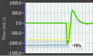
*the above image shows a measured PEFR of 50%, even if the calculated value is 50% or greater the operator can still adjust the t-Low setting to obtain a T-PEFR of up to 75% to maximize lung recruitment. 


Improving Oxygenation: increasing p-High or p-High & t-High

Another way to improve oxygenation during APRV is to increase mean airway pressure. 
One way is to increase p-High to recruit aveoli by achieving threshold opening pressure, p-High should be adjusted at only 2-5 cmH2O increments while monitoring the patients hemodynamic status. 
Furthermore, t-High can be lengthened, this increases gas mixing & recruits alveoli with longer time constants.

note- always assess hemodynamics if increasing p-High or t-High. 
If increasing settings is limited due to decreased cardiac output or hypotension, consider therapeutics which increase cardiac output & blood pressure.

Tuesday, March 24, 2009

Optimal t-Low settings

Optimal t-Low settings are displayed in the images to the left.


 T-PEFR at 50, 60, & 75 percent of the peak expiratory flow rate. 

Notice these precise adjustments of t-Low. 


Friday, March 20, 2009

t-Low: APRV initial settings


Initial t-Low settings are 0.2-0.8 seconds in adult & pediatric patients, and 0.2-0.4 seconds in neonates. 
This short time interval is needed to minimize lung de-recruitment, a longer release phase may interfere with oxygenation. 

note- this cannot be accomplished when using Bi-level on the PB 840 ventilator, where t-Low is not directly set. However, one can manipulate the frequency & "TH" settings to set a t-Low. 

Since atelectasis can develop rapidly when peak airway pressure drops below the opening pressure of the injured lung, t-Low should always be titrated based on analyzing the 'Peak Expiratory Flow Rate' (PEFR) and the 'Peak Expiratory Flow Rate Termination' (T-PEFR) point, utilizing the flow waveform pattern. 

*The above image identifies the PEFR & the T-PEFR in the flow waveform pattern (purple). 


Titration of t-Low

t-Low should be titrated to obtain a T-PEFR at 50-75 percent of measured PEFR.

Example: the above image shows a measured PEFR of -1050 milliliters/second (blue arrow) and a T-PEFR of -350 milliliters/second (yellow arrow). 
This equals 33% of the PEFR
In this example the t-Low would have to be set lower (shorter time) to achieve the 50-75% range to allow for optimal paCO2 removal and maintain enough expiratory lung volume to prevent alveolar de-recruitment.

Observation of the flow waveform should be paired with patient ventilator assessments to identify any need for t-Low adjustments. Changes in pulmonary dynamics result in the need to adjust t-Low settings.


Thursday, March 19, 2009

p-Low: APRV initial settings


To maximize PaCO2 removal p-Low must be set at zero cmH2O. This allows for a rapid peak expiratory flow, which creates a venturi effect that help drafts paCO2 removal. 
Additionally, the high peak expiratory flow rate promptly ends the t-Low phase and allows for the p-High phase to be resumed earlier to optimize alveolar recruitment and exposure time. 

If p-Low is set greater than zero, the peak expiratory flow rate will be decreased and delayed.
Furthermore, a p-Low setting higher than zero creates additional expiratory resistance during the release phase resulting in a more turbulent expiratory flow pattern and decreases the venturi effect.

note- even though p-Low is set at zero lung derecruitment is not a issue, for t-Low is titrated to maintain expiratory lung volume. 
Notice the above image showing that during p-Low there is still pressure in the airways above zero, even when the ventilator is set with a p-Low at 0 cmH2O.